Anatomy Resource Center (ARC)
Dissection lab
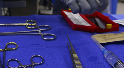
Cadaveric dissection is the most important learning tool to learn gross anatomy. Visualization of actual 3D orientation of the structures is essential to build true concepts of human body. Dissection facility at Alfaisal university can accommodate 10 cadaveric dissection simultaneously. Each student gets ample opportunity to dissect. Dissection of animal organs (e.g. kidney, heart etc.) helps in identifying real texture and improve dissection skills. In near future, gradually formalin embalmed cadaver will be replaced with theil embalmed cadavers which preserves structural detail much better. We strictly follow the Anatomy Act 1984 and the Human Tissue Act 2004.
Dry lab
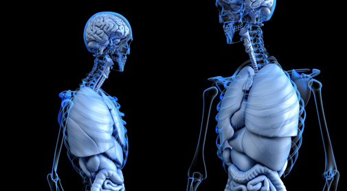
Gross anatomy dry lab has an excellent collection of plastic models, plastinated pro-sections and mounted body sections. These specimens gives an opportunity of 3D visualization and are important in developing basic concept of structure organization. This lab is freely accessible for students during self-directed learning sessions.
Histology Laboratory
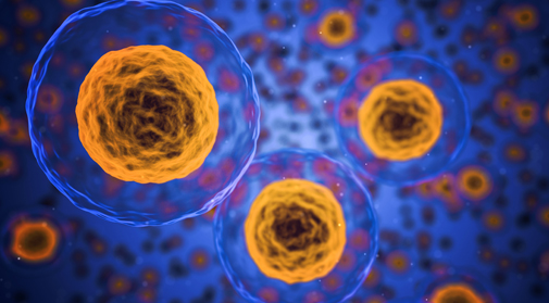
The Histology Laboratory is a combined teaching and research facility supervised by skilled and experienced faculty and lab technicians. It comprises of latest in design light microscopes with finest features. A regularly maintained extensive collection of histology slide and high resolution image library are central to teaching the normal microscopic appearance of the cells and tissues of the body undergraduate students. Histology lab also houses multimedia microscope and projector screens, which are the key resources in delivering of computer assisted lessons. Multimedia microscopes also contribute to the development of a high resolution digital image library essential for teaching, learning and assessment purpose. Lab is also equipped with the latest and advanced high resolution light microscope (BX53F) primarily to assist in graduate program teaching and research activities.
Digital Anatomy Lab (DAL)
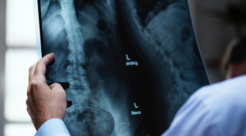
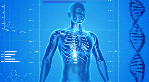
Newly develop Digital Anatomy lab is equipped with state of art 3D virtual dissection tables. These digital tables are loaded with high resolution 3D images with multi view planner. This offers life-size virtual reality which is really helpful in developing 3D conceptualization of human structure. Integrated high resolution radiological images helps in application of basic knowledge. For more clinical integration, soon, Lab will be equipped with ultrasound facility for real life teaching.


